Vision
Our vision has such a large dynamic range that we can adjust to see under the glaring light of high noon to traverse a desert or detect the small scattering of photons that reach our eye after traveling for 2.5 million light-years from the Andromeda Galaxy. This is possible because we have a duplex retina in which signaling from rods in very dim light flawlessly transitions to mixed rod and cone signaling to purely cone signaling as the light intensity increases.
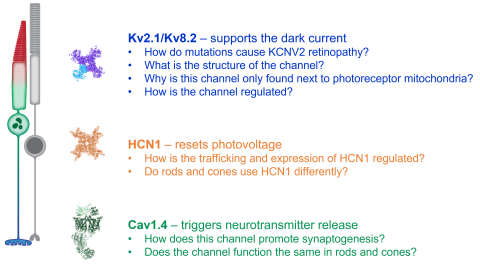
Another factor to the amazing capacity of our visual system is the unique cellular organization of our photoreceptors where a division of labor ensures optimal function. The phototransduction cascade, one of the best studied G-protein signaling pathways, is confined to the membrane discs of the outer segment; while energy production, metabolism, lipid, and protein synthesis are confined to the inner segment. In addition to these housekeeping functions, the inner segment expresses regulatory ion channels that shape the overall response to light. At the opposite end of the cell, ribbon synapses mediate communication to downstream neurons in the retina.
Over the past decade we have been largely focused on studying the role of individual ion channels in photoreceptors. We are especially interested in the biochemical regulation of ion channels and how they are used in rod versus cone signaling. We also seek molecular diagnosis for Inherited Retinal Degenerations. Our work helps the larger vision research community become better positioned to combat blindness.
The Ion Channels Studied by the Baker Lab:
